Novel Application of Indocyanine Green Fluorescence Imaging for Real-Time Detection of Thrombus in A Membrane Oxygenator.

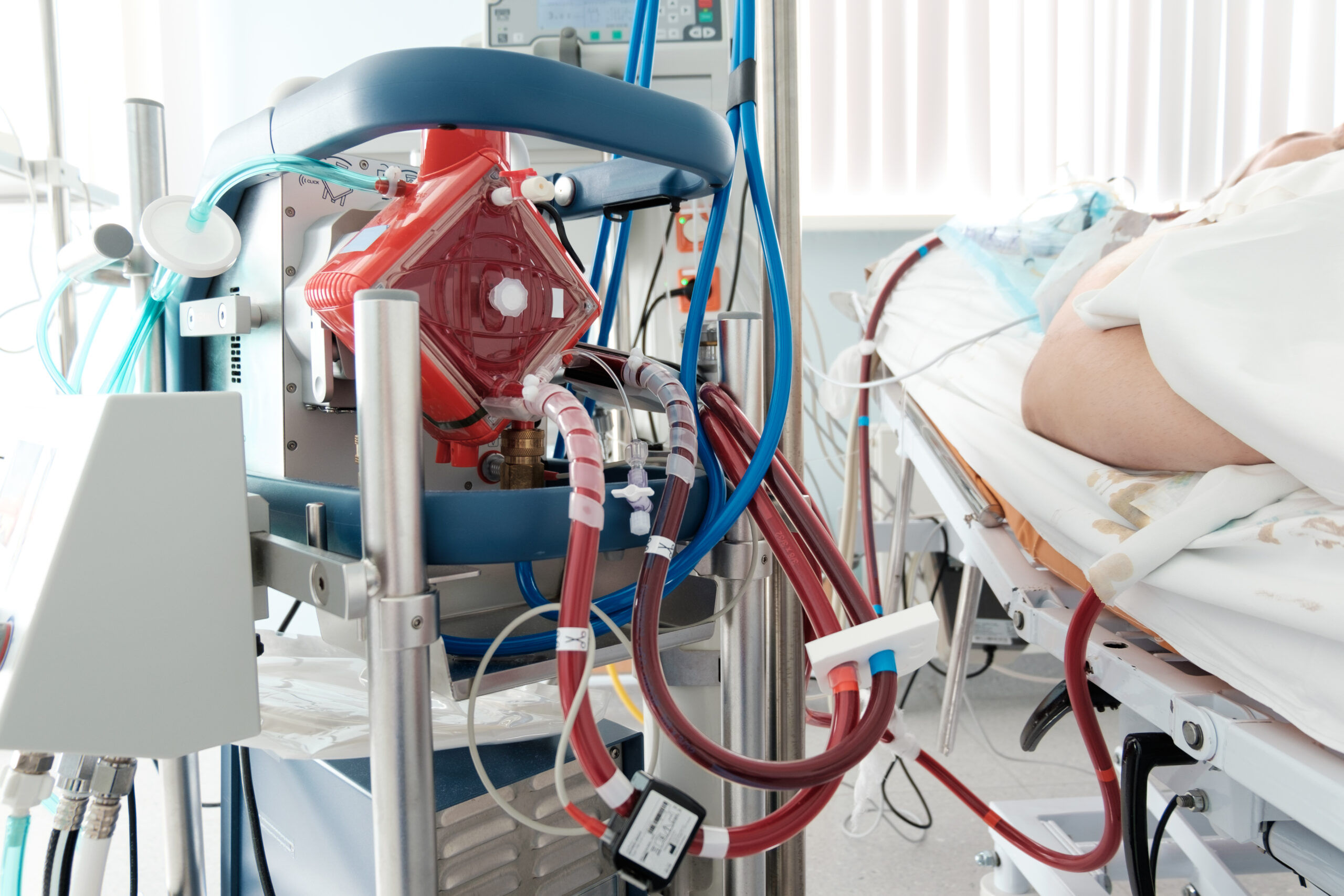
Extracorporeal membrane oxygenation (ECMO) plays an important role in the coronavirus disease 2019 (COVID-19) pandemic. Management of thrombi in ECMO is generally an important issue; especially in ECMO for COVID-19 patients who are prone to thrombus formation, the thrombus formation in oxygenators is an unresolved issue, and it is very difficult to deal with. To prevent thromboembolic complications, it is necessary to develop a method for early thrombus detection. We developed a novel method for detailed real-time observation of thrombi formed in oxygenators using indocyanine green (ICG) fluorescence imaging. The purpose of this study was to verify the efficacy of this novel method through animal experiments. The experiments were performed three times using three pigs equipped with veno-arterial ECMO comprising a centrifugal pump (CAPIOX SL) and an oxygenator (QUADROX). To create thrombogenic conditions, the pump flow rate was set at 1 L/min without anticoagulation. The diluted ICG (0.025 mg/mL) was intravenously administered at a dose of 10 mL once an hour. A single dose of ICG was 0.25mg. The oxygenator was observed with both an optical detector (PDE-neo) and the naked eye every hour after measurement initiation for a total of 8 hours. With this dose of ICG, we could observe it by fluorescence imaging for about 15 minutes. Under ICG imaging, the inside of the oxygenator was observed as a white area. A black dot suspected to be the thrombus appeared 6-8 hours after measurement initiation. The thrombus and the black dot on ICG imaging were finely matched in terms of morphology. Thus, we succeeded in real-time thrombus detection in an oxygenator using ICG imaging. The combined use of ICG imaging and conventional routine screening tests could compensate for each other’s weaknesses and significantly improve the safety of ECMO.
