STS 2.73 : New Risk Factors

The Great Migration to v 2.73 :
(New STS Fields)
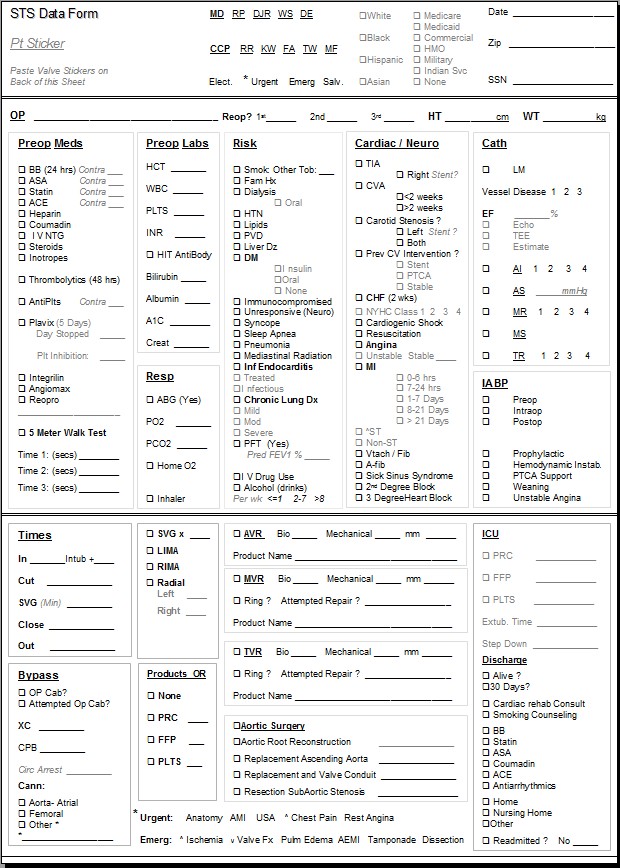
Click Image to View entire 2.73 Series …
The following post describes the new fields that are identified as new data collection points in the STS v. 2.73 Adult Cardiac Database.
(Customized PowerPoint Template for Perfusion STS Data Collection)
- Download
Customized STS Form (for Perfusion)
Overall assessment:
For the Risk Section, some of these fields mimic or build on prior similar field from version 2.61.
Several of these fields (room air ABG, PFT, and the 5-meter walk test- for example) seem to be optional based on the language STS uses in their definition.
It seems that liver function and respiratory function are more significantly emphasized in this version.
The MELD Score in particular (and the components associated with it) comprises an interesting insight to predictive mortality when assessing patient liver function.
Fields marked with an asterisk (*):
*Identifies from a clinical point of view, data that improves or enhances our overall picture of our patient population.
The New STS Risk Fields
* Other Tobacco
Captures use of any tobacco product other than cigarettes within one year.
Includes cigars, pipes, and chewing tobacco. For chewing tobacco, dipping tobacco, snuff, which are held in the mouth between the lip and gum, or taken in the nose, the amount of nicotine released into the body tends to be much greater than smoked tobacco.
Nicotine has very powerful effects on arteries throughout the body. Nicotine is a stimulant, it raises blood pressure, and is a vasoconstrictor, making it harder for the heart to pump through the constricted arteries. It causes the body to release its stores of fat and cholesterol into the blood.
* Platelets
Capture the platelet count at the date and time closest to surgery but prior to anesthetic management.
The administration of IV fluids in the holding area can cause dilution of blood. Do not capture labs drawn after the patient receives fluids in the holding area or O.R.
* INR
Capture the INR at the date and time closest to surgery but prior to anesthetic management.
INR is the standard unit used to report the result of a prothrombin (PT) test. An individual whose blood clots normally and who is not on anticoagulation should have an INR of approximately 1. The higher the INR is, the longer it takes blood to clot.
* HITAntiboby
Indicate whether Heparin Induced Thrombocytopenia, HIT, is confirmed by antibody testing.
Heparin induced thrombocytopenia (HIT) can be defined as any clinical event best explained by platelet factor 4 (PF4) ⁄ heparin-reactive antibodies (‘HIT antibodies’) in a patient who is receiving, or who has recently received heparin. Thrombocytopenia is the most common ‘event’ in HIT and occurs in at least 90% of patients, depending upon the definition of thrombocytopenia. A high proportion of patients with HIT develop thrombosis. Alternative (nonheparin) anticoagulant therapy reduces the risk of subsequent thrombosis.
* TotBlrbn
Indicate the total Bilirubin closest to the date and time of surgery, prior to anesthetic management.
Bilirubin testing checks for levels of bilirubin — an orange-yellow pigment — in blood. Bilirubin is a natural byproduct that results from the normal breakdown of red blood cells. As a normal process, bilirubin is carried in the blood and passes through the liver.
Too much bilirubin may indicate liver damage or disease.
* TotAlbumin
Indicate the albumin level closest to the date and time of surgery, prior to anesthetic management.
Albumin, produced only in the liver, is the major plasma protein that circulates in the bloodstream. Albumin is essential for maintaining the oncotic pressure in the vascular system. A decrease in oncotic pressure due to a low albumin level allows fluid to leak out from the interstitial spaces into the peritoneal cavity, producing ascites. Albumin is also very important in the transportation of many substances such as drugs, lipids, hormones, and toxins that are bound to albumin in the bloodstream.
A low serum albumin indicates poor liver function. Decreased serum albumin levels are not seen in acute liver failure because it takes several weeks of impaired albumin production before the serum albumin level drops. The most common reason for a low albumin is chronic liver failure caused by cirrhosis.
* A1cLvl (for ALL patients not just Diabetics)
Capture the pre-op HbA1c level closest to the date and time prior to surgery.
Glycosylated hemoglobin, HbA1c, is a form of hemoglobin used primarily to identify the average plasma glucose concentration over prolonged periods of time. It is formed in a non-enzymatic glycation pathway by hemoglobin’s exposure to plasma glucose. Normal levels of glucose produce a normal amount of glycosylated hemoglobin. As the average amount of plasma glucose increases, the fraction of glycosylated hemoglobin increases in a predictable way. This serves as a marker for average blood glucose levels over the previous months prior to the measurement.
The 2010 American Diabetes Association Standards of Medical Care in Diabetes added the A1c ≥ 6.5% as a criterion for the diagnosis of diabetes.
* MELDScr
Calculated by software
MELD is a validated liver disease severity scoring system that uses laboratory values for serum bilirubin, serum creatinine and the INR to predict survival.
In patients with chronic liver disease, an increasing MELD score is associated with increasing risk of death.
- ≤ 15 predictive of 95% survival at 3 months
- ~ 30 predictive of 65% survival at 3 months
- ≥ 40 predictive of 10-15% survival at 3 months
MELD = 3.8[Ln serum bilirubin (mg/dL)] + 11.2[Ln INR] + 9.6[Ln serum creatinine (mg/dL)] + 6.4. Laboratory values of INR, total bilirubin and serum creatinine that are <1.0 are set to 1.0. In addition, serum creatinine levels >4.0 mg/dL are capped at 4.0 mg/dL, and patients on dialysis receive an assigned serum creatinine value of 4.0 mg/dL
www.mayoclinic.org/meld/mayomodel6.html
PFT
Indicate whether pulmonary function tests were performed.
This does not imply PFTs should be performed on all patients.
Pulmonary function testing is a valuable tool for evaluating the respiratory system, representing an important adjunct to the patient history, various lung imaging studies, and invasive testing such as bronchoscopy and open-lung biopsy.
- FEV1
If PFTs were done, code the results of FEV1 on the most recent test prior to surgery.
FEV1 is the maximal amount of air forcefully exhaled in one second. It is then converted to a percentage of normal. For example, the FEV1 may be 80% of predicted based on height, weight, and race. FEV1 is a marker for the degree of obstruction with diseases such as asthma In normal persons, the FEV1 accounts for the greatest part of the exhaled volume from a spirometric maneuver and reflects mechanical properties of the large and the medium-sized airways.
•FEV1 > 75% of predicted= normal
•FEV1 60% to 75% of predicted = Mild obstruction
•FEV1 50% to 59% of predicted = Moderate obstruction
•FEV1 < 50% of predicted = Severe obstruction
DLCO
Indicate whether a lung diffusion test was done. (DLCO)
The diffusing capacity (DLCO) is a test of the integrity of the alveolar-capillary surface area for gas transfer.
DLCOPred
Code the results- % predicted of DLCO on the most recent test prior to surgery.
The diffusing capacity (DLCO) may be reduced, <80% predicted, in disorders such as emphysema, pulmonary fibrosis, obstructive lung disease, pulmonary embolism, pulmonary hypertension and anemia.
DLCO>120% of predicted may be seen in normal lungs, asthma, pulmonary hemorrhage, polycythemia, and left to right intracardiac shunt.
ABG
Indicate whether a room air arterial blood gas was performed prior to surgery.
This does not imply all patients should have blood gasses performed.
- PO2
If ABGs were done, indicate the PO2 (partial pressure of oxygen) level on the most recent room air arterial blood gas.
- PCO2
Indicate the PCO2 (partial pressure of carbon dioxide) level on the most recent room air arterial blood gas.
* HmO2
Indicate whether the patient uses supplemental oxygen at home
Capture patients with home oxygen therapy prescribed, despite the amount or frequency of use.
* BDTx
Indicate whether inhaled and/or oral bronchodilator therapy or inhaled (not oral or IV) steroid medications were in use by the patient prior to this procedure.
Capture patients with prescribed bronchodilator therapy, despite amount or frequency of use. Capture prn and routine use.
* SlpApn
Indicate whether patient has a diagnosis of sleep apnea and has been prescribed BiPAP (Bi-level Positive Airway Pressure) or CPAP therapy.
* LiverDis
Indicate whether the patient has a documented history of liver disease.
Liver diseases such as hepatitis B, hepatitis C, cirrhosis, portal hypertension, esophageal varices, chronic alcohol abuse and congestive hepatopathy affect the cells, tissues, structures, or functions of the liver.
ImmSupp
Indicate whether immune compromise is present.
This includes, but is not limited to:
- systemic steroid therapy,
- anti-rejection medications
- chemotherapy.
Has the patient been administered any form of immunosuppressive therapy within 30 days of surgery or was the patient prescribed steroids for chronic or long term usage?
UnrespStat
Indicate whether the patient has a history of non-medically induced, unresponsive state within 24 hours of the time of surgery.
Syncope
Indicate whether the patient had a sudden loss of consciousness with loss of postural tone, not related to anesthesia, with spontaneous recovery and believed to be related to cardiac condition.
Capture events occurring within the past one year as reported by patient or observer. Patient may experience syncope when supine.
Cardiac conditions including dysrhythmias and aortic stenosis can cause syncope.
Do not capture remote episodes of syncope unrelated to cardiac conditions.
CVDCarSten
(This was included in prior versions- but more extensive Carotid Hx is now required)
Indicate which carotid artery was determined from any diagnostic test to be more than 79% stenotic.
Choose none, right, left or both. Diagnostic studies may include ultrasound, doppler, angiography, CT, MRI or MRA. If more than one test was performed with different results, choose the highest level of stenosis reported.
IVDrugAb
Indicate whether patient has a history of illicit drug use such as heroin, marijuana, cocaine, or meth, regardless of route of administration.
- Do not include rare historical use.
Alcohol
Specify Alcohol Consumption History
≤1 drink per week (occasional), 2-7 drinks per week (social) or ≥ 8 drinks per week (heavy). This data element is harmonized with the ACC/AHA definitions.
Pneumonia
Indicate whether patient has a recent or remote history of pneumonia.
Pneumonia is an infection of one or both lungs caused by bacteria, viruses, fungi, chemicals or aspiration. It can be community acquired or acquired in a health care setting.
- No – meaning no history of pneumonia
- Recent- pneumonia diagnosis within 1 month of procedure
- Remote – pneumonia diagnosis more than 1 month prior to the procedure.
MediastRad
Indicate whether patient has a history of radiation therapy to the mediastinum or chest wall.
Mediastinal radiation can cause damage to blood vessels, heart valves and lung tissue. Scar tissue caused by radiation therapy can lead to increased bleeding, may make harvesting the internal mammary artery difficult and may interfere with sternal healing.
Cancer
Indicate whether the patient has a history of cancer diagnosed within 5 years of procedure
Capture cancers that require surgical intervention, chemotherapy and or radiation therapy. Do not capture localized cancers such as skin cancers.
Five MWalkTest (meters)
This gait speed test is a measure of frailty in ambulatory patients, a risk factor previously difficult to capture.
- If done, you will need to record at least one time, ideally 3 times that the system will average.
Frailty is a risk factor for surgery that has been difficult to quantify. This simple test quantifies frailty prior to surgery in ambulatory patients.

