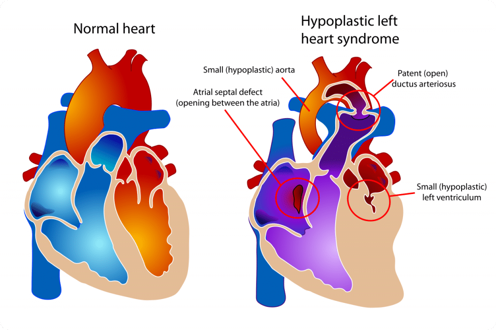
Medical Illustration in the Era of Cardiac Surgery

This article reviews the collaboration between clinician and illustrator throughout the ages while highlighting the era of cardiac surgery. Historical notes are based on Professor Sanjib Kumar Ghosh’s extensive review, literature searches, and the archives of the Johns Hopkins University Department of Art as related to Medicine in Baltimore. Personal communications were explored with medical illustrators and medical practitioners, many of whom are colleagues and trainees, to further chronicle the history of medical illustration and education in the era of cardiac surgery. Medical illustrators use their talents and expressive ideas to demonstrate procedures and give them life. These methods are (1) hovering technique; (2) hidden anatomy, ghosted views, or transparency; (3) centrally focused perspective; (4) action techniques to give life to the procedure; (5) use of insets to highlight one part of the drawing; (6) human proportionality using hands or known objects to show size; and (7) step-by-step educational process to depict the stages of a procedure. Vivid examples showing these techniques are demonstrated. The result of this observational analysis underscores the importance of the collaboration between clinician and illustrator to accurately describe intricate pathoanatomy, three-dimensional interrelated anatomic detail, and complex operations. While there are few data to measure the impact of the atlas on medical education, it is an undeniable assertion that anatomical and surgical illustrations have helped to educate and train the modern-day surgeon, cardiologist, and related health-care professionals.
